Prologue to fluorescence magnifying lens
Albeit a considerable lot of the wonderful disclosures in the field of life science have been made without the use of specific innovation, innovation assumes a significant part in seeing new outskirts in life science. It gives as a way to test and to demonstrate novel thoughts. It additionally gives new data to plan new speculation.
One of wonderful advancements is microscopy and imaging procedures. These strategies involve a middle stage in natural too in material science; a basic apparatus in surveying the properties of natural or inorganic substances. It gives the benefit of having the option to notice and quantify structure and highlights to uncover the inconstancy.
With the improvement of more refine methods couple with the revelation of a variety of fluorophores, it has made conceivable to picture and study the cell and subcellular segments and the assorted physiological cycles like protein connections, particle transports, supplement portability and metabolic cycles occurring inside a living cell.
In light microscopy, the differential reflection, diffraction and retention properties of various examples are utilized to contemplate the examples. So enhancements in microscopy are essentially stressed on expanding the difference between the example and the foundation. This difference between the example and the foundation is improved absolutely by staining the example with specialists that assimilate light in light microscopy.
Anyway this difference has been enormously upgraded with the improvement of fluorescence microscopy. It gives preferable difference over other optical microscopy strategies. This has been accomplished by staining or labeling the objective example with fluorescence color or with fluorescence particles (fluorophores) in the midst of the non-fluorescing foundation. On illumination with high energy light, just the fluorescence atoms transmit light empowering to picture just the object of interest in obscurity foundation.
With the advancement of profoundly explicit marking tests, combined with the capacity to imaging of individual segments and other macromolecular buildings, fluorescence microscopy gets an insurgency cell science.
History of fluorescence
Fluorescence has been depicted first by Irish Scientist Sir George G. Stirs up in 1852 during the center of nineteenth century. While working with a mineral named „fluorspar‟ at the Cambridge University, he saw that the mineral transmitted red light when enlightened with blue light. In spite of the fact that this marvel has been experienced in microscopy in the early piece of 20th century by a few researchers, including August Köhler and Carl Reichert, they neglected to perceived the fluorescence and rather revealed it as a foundation clamor in bright microscopy.
The principal fluorescence magnifying lens were created by German physicists Otto Heimstädt and Heinrich Lehmann somewhere in the range of 1911 and 1913 as variation from the bright microscopy. These magnifying instruments have used to notice autofluorescence in microscopic organisms, creature, and plant tissues. Another period of fluorescence microscopy stirred from that point the improvement of a method for marking antibodies with fluorescent colors by Albert Coons during the mid 1940s.
Excitation and Emission
Each atom can assimilate light of certain frequency. At the point when these atoms are exposed to brilliant energy, they assimilate the energy and become eager to a higher energy state. The intrinsic property of each atom to remain at the most minimal energy state made a big deal about the caught energy to be delivered as warmth or light.
Anyway a few iotas or atoms in the wake of retaining light, it reradiates back the energy as light inside nanoseconds after ingestion. This actual wonder is known as fluorescence and is first depict d by Sir George G. Stirs up. He begat the expression "fluorescence" named after the mineral „fluorspar‟ he is working with.
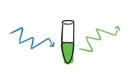
He additionally called attention to that, the light radiated by an energized atom have a frequency longer than the frequency of light initially retained. Endless supply of blue light, green light is discharged soon subsequently. Green is changed to yellow, yellow to rosy orange and undetectable UV light to obvious light. The time delay between the ingestion and outflow of the light in fluorescence is not exactly a microsecond. Nonetheless, the wonder glow happens when the discharge continues even after the excitation light has been suspended.
A helpful clarification of the different energy levels associated with excitation and outflow measure by a fluorophore is outlined by the Jablonski energy graph named after the clean physicist Alexander Jablonski.
At the point when a particle in their ground state assimilates light energy, all the energy of the photon is moved to the atom. The measure of energy retain by the atom is contrarily relative to the frequency of the photon
E=h X c/λ, Where h is the plank‟s steady c and λ are the speed and frequency of light in vacuum.
Subsequent to engrossing the energy the atom is moved to S1 State. Nonetheless if the measure of energy assimilate is more noteworthy than that needs to moved to the S1 state, than the atom to moved to the higher S2 state.
The energized particle remained at the energized state for the period on request of nanoseconds and re-visitation of the ground state by losing the ingest energy. Electrons energized at a higher orbital state (S2) first re-visitation of the S1 state. This change between electron orbitals is known as internal conversion‟.
The additional energy from this progress is lost through vibrational unwinding. Electrons at the S1 state than re-visitation of the ground state. In the event that the drop back from higher energy state S1 to bring down state is joined by the arrival of photon of light whose energy is equivalent to the energy hole between the ground S0 and first single state S1. This radiated light is the fluorescence.
Excitation and Outflow Spectra and Stirs up Shift
A Fluorophore on enlightenment retains light energy. In the event that the energy assimilated is more prominent than that is needed to energize it orbital electrons to S1 state, at that point electrons are moved to the S1 state.
This proposed that a scope of frequency with energies more than the base required moving the orbital electrons to the higher energy state S1. So the excitation range of a fluorophore is the scope of frequencies which can energize the fluorophore. When energized the fluorophore re-visitation of the ground state by arrival of photons of energy. The produced light can be at various frequencies. This scope of frequencies the discharged photon have is the outflow spectra of the fluorophore.
The transmitted spectra is at a more drawn out frequency than the excitation spectra. This distinction between the energizing and radiated frequencies is named as Stoke‟s move named after the Sir George G. Stirs up. This move in frequencies might be ascribed to the energy misfortune because of vibrational and inside transformations that happens by the energized electrons.
Despite the fact that there is a move between the excitation and emanation frequencies, the excitation and outflow spectra are regularly the perfect representation of the other. The excitation and emanation spectra of fluorophore can be controlled by enlightening the fluorophore with various frequencies and estimating the frequencies of light transmitted.
Fluorescence Magnifying Lens
This distinction in frequency between the retention and outflow frequency or Stoke‟s move is the basic property that is being used in fluorescence microscopy; enlightening the test with one frequency and sifting the radiated light to permit just the more drawn out frequency to envision consequently making to see just the articles that are fluorescent against the dull foundation. The figure underneath speaks to the light pathway in an epifluorescence magnifying lens.


0 Comments
We are pleased to see you here! Please mention your suggestion or query in the comments box below.